ResultsĮighty-one infants with a median (range) gestational age of 28.7 (22.4–41.9) weeks were recruited. Alveolar ventilation ( V A) was also calculated. Volumetric capnograms were constructed to calculate the dead space using the modified Bohr–Enghoff equation. Expiratory tidal volume and carbon dioxide levels were measured. MethodsĪ prospective study of mechanically ventilated infants was undertaken.

We determined if there were differences in dead space and alveolar ventilation in ventilated infants with pulmonary disease or no respiratory morbidity.

Positive pressure ventilation (i.e.Dead space is the volume not taking part in gas exchange and, if increased, could affect alveolar ventilation if there is too low a delivered volume.Neck extension and jaw protrusion (can increase it twofold).General anesthesia – multifactorial, including loss of skeletal muscle tone and bronchoconstrictor tone.The ratio of physiologic dead space to tidal volume is usually about 1/3.

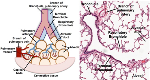
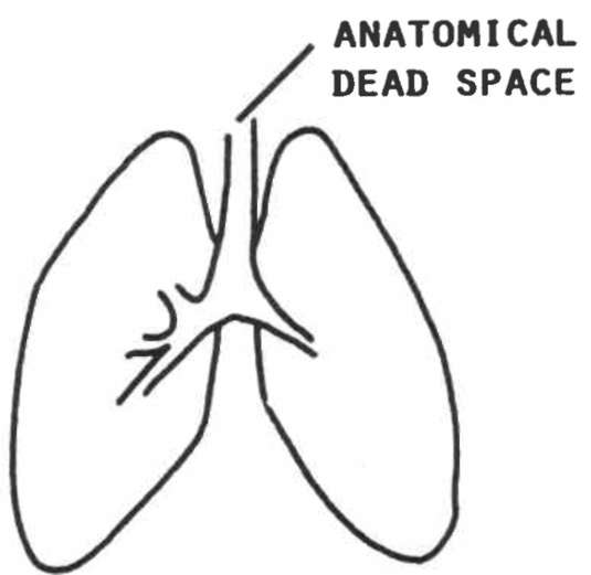
Alveolar dead space is the volume of gas within unperfused alveoli (and thus not participating in gas exchange either) it is usually negligible in the healthy, awake patient. Anatomic dead space is the volume of gas within the conducting zone (as opposed to the transitional and respiratory zones) and includes the trachea, bronchus, bronchioles, and terminal bronchioles it is approximately 2 mL/kg in the upright position. Physiologic or total dead space is the sum of anatomic dead space and alveolar dead space. Dead space is the volume of a breath that does not participate in gas exchange.


 0 kommentar(er)
